Research
4D Breast CT
Breast CT is a fully 3D, isotropic, high spatial and contrast resolution x-ray-based modality which brings the characteristics of tomography to breast imaging. Given these advantages, breast CT has the potential for being used in multiple breast imaging applications, such as detection, diagnosis, staging, and therapy response monitoring. However, clinical performance based upon anatomic evaluation alone is limited due to the similar attenuation of normal fibroglandular and cancerous tissue. Functional information can be acquired via non-invasive imaging by use of a contrast medium and evaluation of its uptake within a patient breast, and especially by lesions. Dynamic uptake information is extremely valuable not only for detection, but especially for lesion classification, reducing the need for biopsies of benign lesions, and for treatment planning and response monitoring.
In this project we aim at development and optimization of 4D dynamic contrast-enhanced breast CT, a modality able to provide four-dimensional images (i.e. 3D over a continuous time range) of breast cancer. Thanks to its physical unique properties, 4D breast CT is envisioned to achieved an improved spatio-temporal characterization of breast cancer, potentially resulting in strong imaging biomarkers that could be used to improve diagnosis and guide therapy.
The 4D breast CT system will be designed through the development of digital phantoms, computer simulations, and algorithms for image denoising and motion and scatter correction, with the goal of optimizing this technique in terms of image quality and delivered radiation dose. Subsequently, radiomics algorithms will be developed and applied to quantify the cancer imaging biomarkers in the 4D images, and used to develop models for clinical decision support.
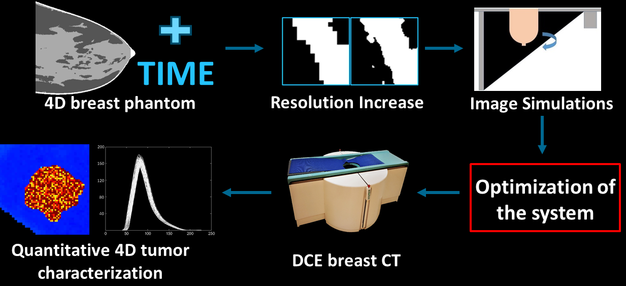
Main pipeline for the 4D breast CT project. 4D digital breast phantoms which can realistically mimic the perfusion patterns of different tissues and lesions are being developed, and their spatial resolution is increased through Machine Learning to generate super-resolution realistic breast models. These models will be used for computer simulations of 4D breast CT, to optimize this imaging modality in terms of image quality and delivered radiation dose. The final steps include the development and validation of an image analysis algorithm for the automatic extraction and quantification of functional tumor-related imaging biomarkers, which could be used to improve the diagnosis and guide the therapy.
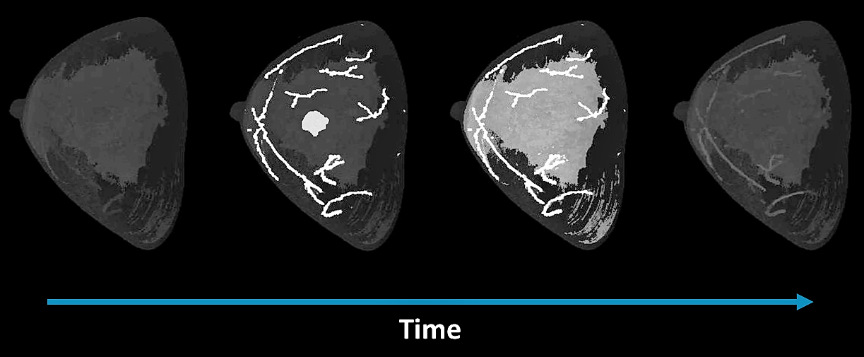
Dedicated digital breast phantom simulating functional behaviour and Iodine uptake in a breast, to be used as input for the entire 4D breast CT simulation chain.
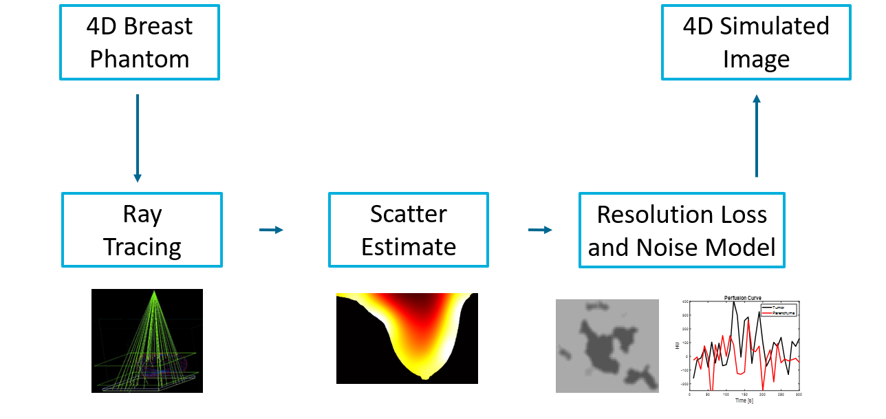
Computerized image simulation pipeline for 4D perfusion breast CT, which can be used to investigate the effect of different hardware components and acquisition settings on the resulting image quality.
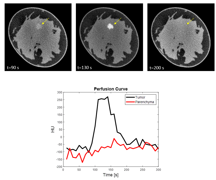
Example of simulated 4D breast CT image.
Researchers:
Key Publications:
- M. Caballo, K. Michielsen, C. Fedon and I. Sechopoulos. "Towards 4D dedicated breast CT perfusion imaging of cancer: development and validation of computer simulated images.", 2019. Abstract. DOI.
- M. Caballo, J. Boone, R. Mann and I. Sechopoulos. "An unsupervised automatic segmentation algorithm for breast tissue classification of dedicated breast computed tomography images.", 2018. Abstract. DOI.
- M. Caballo, C. Fedon, L. Brombal, R. Mann, R. Longo and I. Sechopoulos. "Development of 3D patient-based super-resolution digital breast phantoms using machine learning.", 2018. Abstract. DOI.
- M. Caballo, R. Mann and I. Sechopoulos. "Patient-Based 4D Digital Breast Phantom for Perfusion Contrast-Enhanced Breast CT Imaging.", 2018. Abstract. DOI.
- M. Caballo, C. Fedon, L. Brombal, K. Michielsen, R. Longo and I. Sechopolous. "Automatic estimation of glandular tissue loss due to limited reconstruction voxel size in tomographic images of the breast", 2018. Abstract. DOI.
- J.J. Pautasso, M. Caballo, M. Mikerov, J.M. Boone, K. Michielsen, I. Sechopoulos. "Deep learning for x-ray scatter correction in dedicated breast CT." 2023. Abstract. DOI.
- J.J. Pautasso, K. Michielsen, I. Sechopoulos. "Technical note: Characterization, validation, and spectral optimization of a dedicated breast CT system for contrast-enhanced imaging". 2023. Abstract. DOI.
- J.J. Pautasso, M. Mikerov, L. Goris, K. Michielsen, I. Sechopoulos. "4D dynamic contrast-enhanced breast CT: evaluation of quantitative accuracy". 2024. Abstract. DOI.