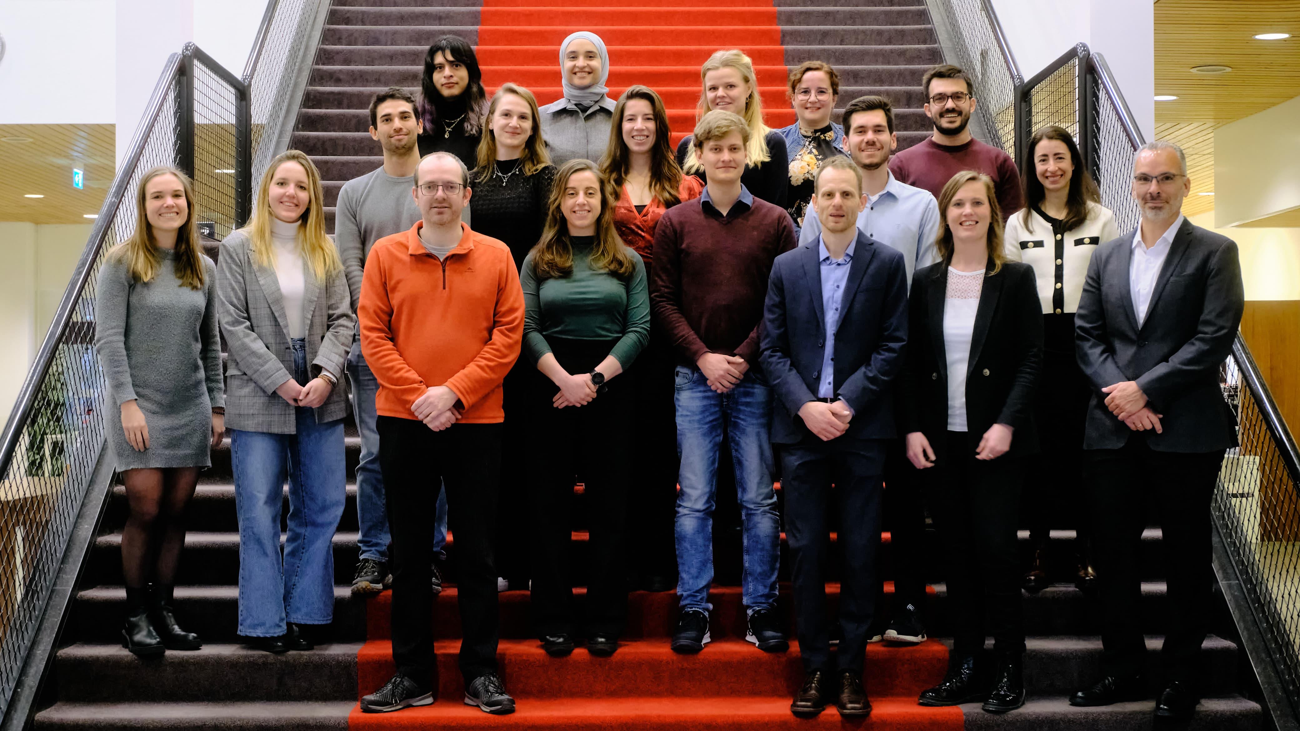
The Advanced X-ray Tomographic Imaging (AXTI, homophone of actie, meaning action in Dutch) lab specializes on research on novel image acquisition, processing and analysis. Its scope include the physical and perceptual evaluation of image quality, radiation dosimetry, tomographic image reconstruction and advanced computational simulation methods. This research primarily focuses on diagnostic x-ray based imaging, particularly for breast and body tomographic.
The AXTI lab aims to conduct state-of-the-art and clinically relevant research through collaboration with clinicians from the Department of Medical Imaging at Radboudumc and partnerships with commercial entities, medical physicists, and scientists from around the world.
With a team possessing diverse skills and expertise, AXTI's research encompasses the entire medical imaging chain, from image generation physics to optimizing image interpretation by both radiologists and AI-based algorithms. Projects include developing advanced tomographic breast imaging methods for cancer detection, diagnosis, and personalized treatment. Research also focuses on non-invasive diagnosis of ligament damage using functional dynamic CT of the wrist, and enhancing breast cancer screening with mammography, including AI-supported interpretation.
AXTI is part of Radboud Research Institute's “Breast Cancer” and the “Imaging technologies for diagnosis, treatment, and elucidating pathophysiology of diseases” Research Programs. Funding comes primarily from the Netherlands Organisation for Scientific Research (NWO), the European Research Council (ERC), the Dutch Cancer Foundation (KWF), and others. Collaborations include Siemens Healthineers, Canon Medical Systems, and Screenpoint Medical, and with the Dutch Expert Centre for Screening (LRCB).
