Research
Breast Shape Models
Many applications in breast imaging such as dosimetry models, x-ray scatter correction algorithms, and image processing techniques require realistic virtual breast models. We have created realistic 3D models of the shapes of compressed breasts undergoing mammography or breast tomosynthesis for this use, in both the cranio-caudal (CC) and medio-lateral oblique (MLO) view.
A protocol was developed to accurately capture the 3D shape of patients’ breast before and during compression for mammography. Using a pair of 3D surface scanners, the patient breasts are scanned, resulting in accurate representations of the breast surfaces during mechanical compression by the mammography system. From this data, we developed a computer model that can generate a representative sample of 3D compressed breast shapes, with potential application in the improvement of digital breast tomosynthesis image reconstruction, and in the validation of computer algorithms that simulate mechanical deformations of the breast.
The end goal for this latter application is to create and validate an image processing algorithm able to track the tumor position from x-ray images for different breast positions (e.g. prone and supine position), with the improvement in surgical planning as a potential outcome.
Due to the success of the initial studies, we have started a new collaboration with Siemens Healthineers on the development of a more polished prototype. We are currently working on calibration and alignment of the sensors. These sensors will be used to investigate the benefit of performing patient-specific image processing.
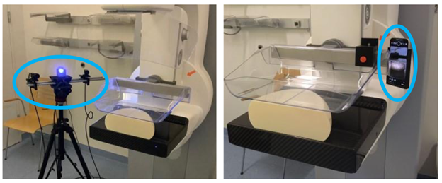
Camera setup of the structured light scanning system used for the CC view (left), locally mounted smartphone-based infrared scanning system used for the MLO view (right), with a phantom placed on the breast tomosynthesis system.
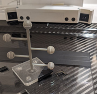
Custom alignment fiducials used to calibrate the new sensors in collaboration with Siemens Healthineers. The spheres are made from specialized infrared reflective material to aid in separating the relevant data from background noise.
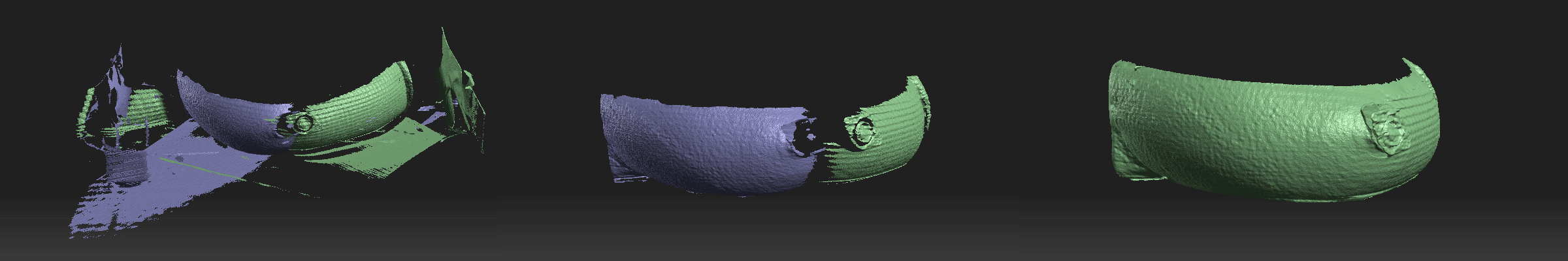
Post processing steps of the 3D external breast surface, from left to right: Raw surface images acquired by the two surface cameras; cleaned images with signals that do not belong to the breast surface removed; and merged surfaces from both lateral cameras after fusion using global fine registration.
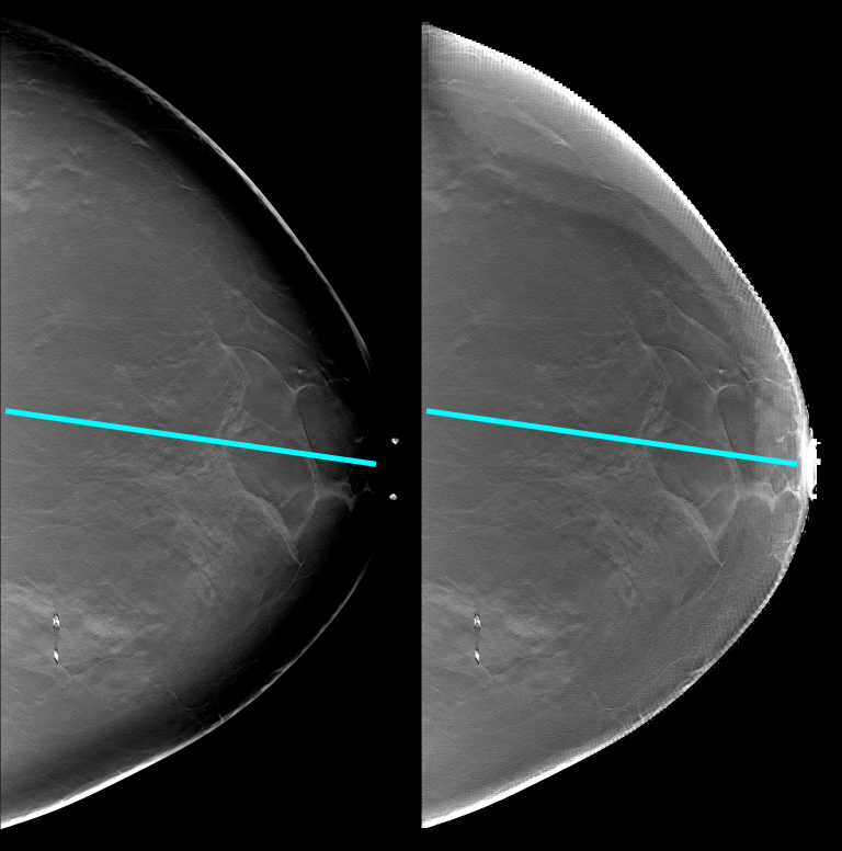
These two images show the difference between a standard tomosynthesis reconstruction (left) and a reconstruction which includes the information from the surface scan of the compressed breast (right). Adding this information recovers the attenuation values near the breast edge and allows for better visualization of that region.
Researchers:
Key Publications:
- Pinto, M. C., T. H. Egten, K. Michielsen, R. Biniazan, S. Kappler, and I. Sechopoulos. “First Evaluation of 3D Measurements of the Compressed Breast Shape for Digital Breast Tomosynthesis in the Medio-Lateral Oblique View.” In Medical Imaging 2023: Physics of Medical Imaging, 12463:567–71. SPIE, 2023. DOI.
- Pinto, Marta C., Franziska Mauter, Koen Michielsen, Ramyar Biniazan, Steffen Kappler, and Ioannis Sechopoulos. “A Deep Learning Approach to Estimate X‐ray Scatter in Digital Breast Tomosynthesis: From Phantom Models to Clinical Applications.” Medical Physics 50, no. 8 (August 2023): 4744–57. DOI.
- Pinto, Marta C., Koen Michielsen, Ramyar Biniazan, Steffen Kappler, and Ioannis Sechopoulos. “Generative Compressed Breast Shape Model for Digital Mammography and Digital Breast Tomosynthesis.” Medical Physics 50, no. 5 (May 2023): 2928–38. DOI.
- Pinto, Marta B., Joana Boita, Koen Michielsen, and Ioannis Sechopoulos. “iPhone TrueDepth Cameras Performance Compared to Optical 3D Scanner for Imaging the Compressed Breast Shape.” In 16th International Workshop on Breast Imaging (IWBI2022), edited by Hilde Bosmans, Nicholas Marshall, and Chantal Van Ongeval, 4. Leuven, Belgium: SPIE, 2022. DOI.
- K. Michielsen, A. Rodriguez-Ruiz and I. Sechopoulos. "Estimating the compressed breast-shape using deep learning.", 2020. Abstract. DOI.
- A. Rodríguez-Ruiz, G. Agasthya and I. Sechopoulos. "The compressed breast during mammography and breast tomosynthesis: in vivo shape characterization and modeling.", 2017. Abstract. DOI.