Research
Deep Learning for Image Quality Assessment in Mammography
Regular quality control (QC) of mammography screening devices is of vital importance to ensure optimal and accurate image acquisition. In compliance with regulations, QC measurements are currently carried out with technical phantoms that have little resemblance to a female breast. In one such measurement, using the so-called CDMAM phantom (see Fig. 1) the task of small lesion detection is mimicked. Although measured by a mathematical model observer, the result of this quality measurement should give an estimate of how well a human observer is able to detect small lesions with the tested mammography imaging device. This is an important criterion linking the measure of image quality to the clinical use of the device.
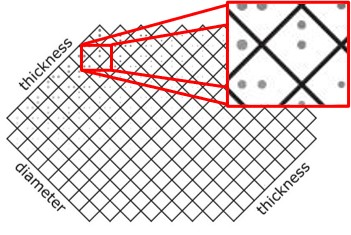
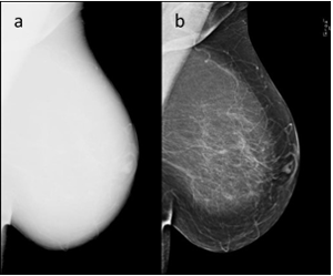
Top: Schematic of the CDMAM phantom. It consists of 205 grid cells with 2 gold discs within each cell varying in thickness or diameter depending on the phantom axis. Bottom: Mammogram of a female breast. a) Unprocessed image “for processing”. b) Processed image “for presentation”.
However, although claiming to be a task-based image quality assessment method, this procedure is neither truly able to replicate the task of small lesion detection in a mammogram, nor is it actually relatable to human observer performance. Since the CDMAM phantom is a technical phantom, it is lacking the anatomy of the female breast. This means that although the linear imaging stages involved in image creation can be tested adequately with it, this is not true for the entire image formation chain. This is because the internal processing software of modern mammography devices alters the original, raw image (“for processing”, see Fig. 2a) using non-linear processes. This makes the images easier to review (the processed image then being called “for presentation”, see Fig. 2b). Unfortunately, this processing software behaves differently with images of real breasts compared to images of technical phantoms and thus this processing step cannot be tested with the CDMAM phantom. However, since data processing can have great impact on the perceived image quality, it should be included in QC tests. The reason for lacking relatability to human observer performance is due to the missing correlation between mathematical model observer and human observer. Another downside of the current procedure using the CDMAM phantom is that it requires several images to be taken so that a quality metric – the contrast-detail curve – can be estimated reliably.
Within this project, we aim to address and resolve these downsides. By using realistic anatomical phantoms, we want to build and train a deep learning observer (DLO) to derive quality metrics from just a few (preferably only one) images (see Fig. 3). These anthropomorphic phantoms would have the advantage of providing the possibility to develop a QC procedure that is more similar to the clinical task of small lesion detection and to include the processing step into the quality measure. Furthermore, using deep neural networks allows for training the DLO to be better relatable to human observers. Finally, deriving a quality metric from only a single image would reduce the time, effort, and cost of QC measurements.
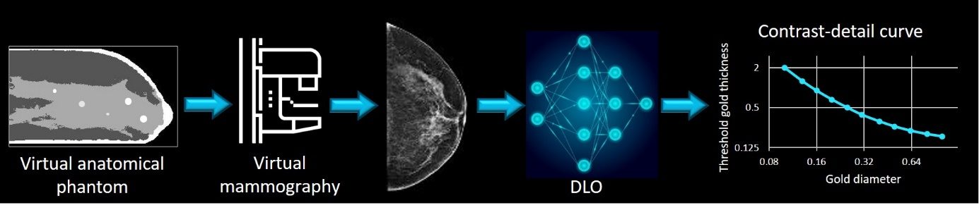
Schematic overview of planned QC procedure. A deep learning observer will be trained on virtually simulated mammograms of anatomical phantoms to determine a measure of image quality (e.g., a contrast-detail curve).
This project is a collaboration between the German National Metrology Institute (PTB) in Braunschweig, Radboud University Medical Center in Nijmegen and the Dutch Expert Center for Screening (LRCB , Nijmegen).
Researchers: